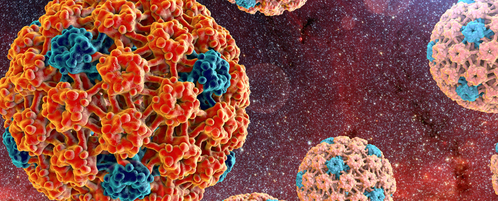
Sexually transmitted infections (STIs) are among the most frequent infectious diseases throughout the world and are defined as infectious organisms which are transmitted between sex partners. According to the Centers for Disease Control (CDC), about 19 million cases are reported each year with more than 20 different STIs (1). Human papilloma virus (HPV) is one of the most frequent causes of STIs in women worldwide with more than 200 different HPV genotypes that are generally classified into high- and low-risk groups based on their potential risk of causing cancer. About 99% of all cervical malignancies are one or more of the following HPV high-risk types: 16, 18, 31, 35, 39, 45, 51, 52, 56, 58, 59. High-risk types also play a role in other cancers including anal, oropharyngeal, vulvar, vaginal, and penile.
HPV is easily transmitted from one person to another via skin and mucus membranes, and while relatively common, the majority of infections are subclinical and temporary due to suppression and clearance by an immunocompetent immune system. Cervical cytology and HPV tests are widely used for cervical cancer screening in countries that have easy access for people, and thus early detection is considered a key aspect of cervical cancer protection in particular.
The human microbiome is the sum of microorganisms that may reside in various parts of the body, their genetic information, and how they interact with the environment of the host. While we now have a significant amount of data mapping the microbiota in several sites of the human body, especially the gut, there is emerging evidence that the vaginal microbiota may play a key role in HPV carcinogenesis(2) and is related to protection against dysbiosis as well as HPV infection (3,4).
In healthy reproductive-aged women, vaginal pH is primarily determined by lactic acid-producing bacteria, primarily lactobacillus species. If lactobacilli do not dominate the vaginal microbiota, a woman’s antibacterial defensive mechanisms are compromised (5). Alterations in vaginal microbiota and respective changes in vaginal pH are associated with bacterial vaginosis, Chlamydia trachomatis, trichomoniasis, and urinary tract infections. Five major community state types (CST) in the vagina have been described (6). Researchers have studied the vaginal microbiota of 396 asymptomatic women and characterized the species into five groups based on their genes. In a healthy vaginal environment, CST I, II, III, and V are dominated by Lactobacillus crispatus, L. gasseri, L. iners and L. jensenii, respectively. CST IV is characterized by depletion of lactobacilli and increased diversity of anaerobic bacteria such as Atopobium (7).
In 2020, an extensive systematic review of studies reporting data on the association of microbiota and HPV was published (1). Of the 78 articles retrieved from PubMed and 291 from Scopus, 16 studies were eligible for inclusion in the review. These 16 studies included a total of 1,204 patients. The detected microbiotas in these studies included several types of microorganisms: L. iners, which is classified as CST III, was found in 13 studies (72.2%); L. crispatus, a CST I classification, was found in 8 studies (44.44%) and CST IV-B, which represents anaerobic microbiomes combined with a reduction in Lactobacillus, was found in 5 studies (27.7%); Megasphaera, Gardnerella vaginalis, and L. jensenii, which are classified as CST V, were found in 4 studies (22.22%); Sneathia and L. gasseri, classified as CST II as well as CST IV-A, representing Peptoniphilus, Anaerococcus, Cornebacterium, Finegoldia and Prevotella, were found in 2 studies (12.5%); and in one study each (6.25%), dialister, L. formicalis, Fusobacterium, L. gallinarum, and L. salivarus (which was found only in South African women).
What does all of this mean regarding vaginal microbiota association with HPV and cervical intraepithelial neoplasia (CIN)? In one of the studies in the review, women with HPV had a higher diversity and a lower proportion of Lactobacillus with a specific lower prevalence of L. iners and L. crispatus. Other common organisms among HPV-positive women were L. gasseri and G. vaginalis. In another study, women who were eventually diagnosed with CIN also had a high diversity of microbiota and were usually colonized by Sneathia, and in women with invasive cervical cancer, Fusobacterium was the most common type of organism. In another study included in the review, there was an abundance of Lactobacillus and L. reuteri specifically in women with CIN II. Contrast that with HPV-negative women who had L. crispatus/CST I and L. gasseri/CST II as the most common species. L. crispatus appears to be related to decreased prevalence of oncogenic HPV types and high-risk HPV infections appear to have a decreased population of Lactobacillus and an increased abundance of anaerobes, particularly Prevotella and Leptotrichia.
HPV remission is another area of interest, and in one report included, CST III was the classification group with the fastest remission, while CST IV-B was the one with the slowest, with CSTIV-B being a risk factor for HPV persistence. Remember from earlier in this article, in a healthy vaginal environment, CST III is dominated by L. iners and CST IV is characterized by depletion of lactobacilli and increased diversity of anaerobic bacteria such as Atopobium. CST IV-A represents Peptoniphilus, Anaerococcus, Corynebacterium, Finegoldia and Prevotella.
Women who are HPV-negative who later become HPV-positive may have a higher CST IV-A microbiota than those with CST I. L. crispatus has another standout feature in that it was found to be a protective factor against HIV, high-risk HPV, and Herpes Simplex type 2, with a high abundance in uninfected women.
Ethnicity is another factor that strongly affects the vaginal microbiota. In this review, Afro-Caribbean women have a fourfold higher risk of suffering from a vaginal dysbiosis or high microbiota diversity, which indicates that CST IV is the most common type of microbiota in comparison with European/Caucasian and African women. Even though this was the case, the prevalence of HPV and the rate of more severe dysplasia was not proportionately higher, which is surprising.
In summary, among all the microbiota, it’s Fusobacteria including Sneathia as possible microbiological markers correlated with HPV although the relationship between HPV infection and the coexistence with other types of vaginal microbiota is either protective or predisposing to HPV. The evolution of HPV infection is in direct correlation with the dominant vaginal species or genus. L. gasseri, L. jensenii, and L. crispatus seem to be protective, while Sneathia, Anaerococcus tetradius, Peptostreptococus, Fusobacterium, G. vaginalis, and L. iners, in combination with a low amount of the other types of Lactobacilli, lead to elevated rates of HPV infection, greater disease severity, and lower rates of HPV remission. Other factors such as nicotine use, lack of barrier contraception, and low vaginal estrogen can also lead to elevated rates of HPV infection. The low vaginal estrogen connection is again related to the vaginal microbiome and the subsequent lower amounts of lactic acid-producing lactobacilli in the postmenopausal state.
Perhaps the greatest limitation of this study and how to put it to clinical use is that we don’t use the tools of DNA tests, sequencing and polymerase chain reaction amplification of genes, gram stains, microbiological cultures, and vaginal pH in our usual assessment and management of HPV. For persistent HPV infections and/or higher-grade lesions with recurrences in particular, we could incorporate at least some of these tests in addition to HPV DNA testing, the most easily being vaginal pH, gram stains, and microbiological cultures. There are vaginal microbiome tests on the market, even some for home use. One is called EVVY. Molecular methods of next generation sequencing are being used to characterize the vaginal microbiota and even a single vaginal swab sample, a nucleic acid amplification test (NAAT), can detect small amounts of microbial DNA and assess overall diversity of the vaginal microbiome.
When it comes to intervention with probiotics and HPV, we are at the early edge of understanding interventions, but particular species, as well as nutraceutical proteins such as lactoferrin, deserve attention. For the moment, our creative thinking in connecting dots and clinical judgement on protocols is in play, perhaps gleaning from some of the research on vaginal probiotics and bacterial vaginosis in terms of dosing regimens. For starters, I’m going to look towards vaginal L. crispatus, L. jensenii, and L. gasseri, along with testing for bacterial vaginosis and the use of vaginal estrogen in postmenopausal women with persistent and/or recurrent HPV/CIN, in terms of influencing the vaginal microbiota. In other NFH resources, for example the Women’s Health Clinical Handbook, we also have data on the use of green tea extract (oral and compounded suppositories), Trametes versicolor, DIM, selenium, and folate for use in abnormal cervical cytology and HPV. I have developed protocols that are available in the handbook.
References:
- Mortaki D, Gkegkes I, Psomiadou V, et al. Vaginal microbiota and human papillomavirus: a systematic review. J Turk Ger Gynecol Assoc 2020; 21:193-200.
- Krygiou M, Mitra A, Moscicki A. Does the vaginal microbiota play a role in the development of cervcal cancer. Transl Res 2017; 179: 168-182.
- An de Wijgert J, Borgdorff H, Verhelst R, et al. The vaginal microbiota: what have we learned after a decade of molecular chaeracterization? PLoS One 2014;9:e105998.
- Brotman R. Vaginal microbiome and sexually transmitted infections: an epidemiologic perspective. J Clin Invest 2011;121: 4610-7.
- Linhares I, Summers P, Larsen B, et al. Contemporary perspectives on vaginal pH and lactobacilli. Am J Obstet Gynecol 2011;204:120.
- Ravel J, Gajer P, Abdo Z, et al. Vaginal mirobiome of reproductive-age women. Proc Natl Acad Sci USA 2011; 108 (Suppl 1): 4680-7.
- Mortaki D, Gkegkes I, Psomiadou V, et al. Vaginal microbiota and human papillomavirus: a systematic review. J Turk Ger Gynecol Assoc 2020; 21:193-200.


